

(1997) Clinical diagnosis of ocular sarcoidosis.FALL 2011 EXECUTIVE OFFICE OF THE SECRETARY-GENERALĮxecutive Office of the Secretary-General NL-03018Ĭhef de Cabinet Under-Secretary-General Nambiar Vijay K. Stavrou, P., Linton, S., Young, D.W., Murray, P.I.

(2006) Cytokines and chemokines in uveitis: Is there a correlation with clinical phenotype?. Ooi, K.G., Galatowicz, G., Calder, V.L., Lightman, S.L. (1995) Branch retinal vein occlusion in a child with ocular sarcoidosis. Ohara, K., Okubo, A., Sasaki, H., Kamata, K. (1978) Sarcoidosis and its ophthalmic manifestations. Obenauf, C.D., Shaw, H.E., Sydnor, C.F., Klintworth, G.K. Sarcoidosis and other granulomatous disorders, New York: Marcel Dekker, 181 (2006) Ocular sarcoidosis: A diagnostic challenge. (1986) Ocular involvement in chronic sarcoidosis. (1997) Global epidemiology of sarcoidosis: What story do prevalence and incidence tell us?. International Ophthalmology Clinics, 47(4): 67 (2007) Neuro-ophthalmologic manifestations of sarcoidosis. However, in patents that are refractory to corticosteroids, methotrexate has shown the most potential as alternative.īraswell, R.A., Kline, L.B. In severe cases, systemic corticosteroid therapy always constitutes the first approach. Recognition of sarcoidosis includes compatible radiological and clinical presentation with histological evidence of noninfectious and noncaseating epitheloid cell granulomas. On average, patients lost 3,4 lines of visual acuity during the follow-up period. In some patients (35 %) regular monitoring may be all that is required, as a significant proportion of patients will show spontaneous improvement. Total exudative detachment of the retina was seen in one eye. One patient had bilateral orbital granulomas that did require treatment. Cystoid macular edema, retinal neovascularisation, disc edema, and optic nerve granulomas also occur. These patients manifested many signs typical of ocular sarcoidosis, including the bilateral nature of the disease, and mutton-fat keratic precipitates, Koeppe and Busacca iris nodules, white clumps of cells (snowballs) in the anterior inferior vitreous, linear or patchy retinal periphlebitis presents as sheathing. There were 15 patients with a mean age at diagnosis of 47 years (range, 10-65) and mean follow-up of 6.1 years (range, 0-15).
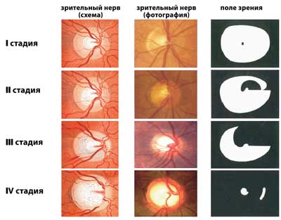
Edem papile skin#
A review of their historical, clinical, laboratory investigations, (hypercalcaemia with hypercalciuria, elevated angiotensin-converting enzyme (ACE), and other diagnostic tools (bronchoalveolar lavage, tuberculin skin test, HIV serological test) and fluorescein angiographic features was made. All patients examined in the authors' referral practices for ocular sarcoidosis diagnosed after the age of 10 were identified. To investigate manifestations and clinical course of ocular sarcoidosis, diagnosed in childhood and adulthood, and to describe characteristics of patients who develop it. Međutim kod reftrakternih slučajeva metotreksat se pokazao kao veoma potentan u tzv. Kod težih slučajeva sistemski kortiko preparati su bili prva linija terapije. Prepoznavanje sarkoidoze podrazumeva radiološku i kliničku dijagnostiku sa histološkom potvrdom neinfektivnog i nekazeoznog granuloma epiteloidnih ćelija. U vremenskom periodu u kom su bili praćeni pacijetni su u proseku gubili 3,4 linije u vidnoj oštrini. Kod 35% pacijenata je bio potreban samo redovan monitoring, i to je bilo u proporciji sa brojem pacijenata koji pokazuju spontano poboljšanje. Totalna eksudativna ablacija je uočena takođe kod jednog pacijenta. Kod jednog pacijenta gde je dijagnostikovan bilateralni orbitalni granulom, bila je potrebna dodatna terapija. Cistoidni makularni edem, neovaskularizacija retine, edem papile, i granulom papile takođe su bili prisutni. Kod ovih pacijentata su uočeni brojne promene tipične za okularnu sarkoidozu, uključujući bilateralnu prirodu bolesti, slaninaste precipitate na endotelu rožnjače, Kepeove i Busaka nodule, snežne lopte u prednjem vitreusu, znake linearnog i 'krpastog' periflebitisa, prisutno kao tzv. (rang, 10-65) sa prosečnim praćenjem 6.1 godine. Ukupno je bilo 15 pacijenata prosečne starosti 47 g. Dat je prikaz kliničke slike sa anamnezom i laboratoratorijom (hiperkaciemija, sa kalciurijom, povišen angiotenzin-konvertujući enzim (ACE)), kao i drugim dijagnostičkim pretragama (bronhoalveolarna lavaža, tuberkulin kožni test, HIV seroloski test) i fluoresceinska angiografija. Svi pacijenti ispitani od strane autora su imali dijagnostikovanu okularnu sarkoidozu i bili su stariji od 10 godina. Prikaz manifestacija i kliničke slike okularne sarkoidoze dijagnostikovane kod odraslih i dece sa opisom karakteristika pacijenata kod kojih se pojavila.


 0 kommentar(er)
